Hip
- Anatomy
- Conditions
- Procedures
Hip Joint
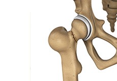
The hip joint is the largest weight-bearing joint in the human body. It is also referred to as a ball and socket joint and is surrounded by muscles, ligaments, and tendons. The thigh bone or femur and the pelvis join to form the hip joint.
Any injury or disease of the hip will adversely affect the joint's range of motion and ability to bear weight.
The hip joint is made up of the following:
- Bones and joints
- Ligaments of the joint capsule
- Muscles and tendons
- Nerves and blood vessels that supply the bones and muscles of the hip
Bones and Joints
The hip joint is the junction where the hip joins the leg to the trunk of the body. It is comprised of two bones: the thigh bone or femur and the pelvis which is made up of three bones called ilium, ischium, and pubis. The ball of the hip joint is made by the femoral head while the socket is formed by the acetabulum. The Acetabulum is a deep, circular socket formed on the outer edge of the pelvis by the union of three bones: ilium, ischium, and pubis. The lower part of the ilium is attached by the pubis while the ischium is considerably behind the pubis. The stability of the hip is provided by the joint capsule or acetabulum and the muscles and ligaments which surround and support the hip joint.
The head of the femur rotates and glides within the acetabulum. A fibrocartilagenous lining called the labrum is attached to the acetabulum and further increases the depth of the socket.
The femur or thigh bone is one of the longest bones in the human body. The upper part of the thigh bone consists of the femoral head, femoral neck, and greater and lesser trochanters. The head of the femur joins the pelvis (acetabulum) to form the hip joint. Next, to the femoral neck, there are two protrusions known as greater and lesser trochanters which serve as sites of muscle attachment.
Articular cartilage is the thin, tough, flexible, and slippery surface lubricated by synovial fluid that covers the weight-bearing bones of the body. It enables smooth movements of the bones and reduces friction.
Ligaments
Ligaments are fibrous structures that connect bones to other bones. The hip joint is encircled with ligaments to provide stability to the hip by forming a dense and fibrous structure around the joint capsule. The ligaments adjoining the hip joint include:
- Iliofemoral ligament: This is a Y-shaped ligament that connects the pelvis to the femoral head at the front of the joint. It helps in limiting the over-extension of the hip.
- Pubofemoral ligament: This is a triangular shaped ligament that extends between the upper portion of the pubis and the iliofemoral ligament. It attaches the pubis to the femoral head.
- Ischiofemoral ligament: This is a group of strong fibers that arise from the ischium behind the acetabulum and merge with the fibers of the joint capsule.
- Ligamentum teres: This is a small ligament that extends from the tip of the femoral head to the acetabulum. Although it has no role in hip movement, it does have a small artery within that supplies blood to a part of the femoral head.
- Acetabular labrum: The labrum is a fibrous cartilage ring which lines the acetabular socket. It deepens the cavity, increasing the stability and strength of the hip joint.
Muscles and Tendons
A long tendon called the iliotibial band runs along the femur from the hip to the knee and serves as an attachment site for several hip muscles including the following:
- Gluteals: These are the muscles that form the buttocks. There are three muscles (gluteus minimus, gluteus maximus, and gluteus medius) that attach to the back of the pelvis and insert into the greater trochanter of the femur.
- Adductors: These muscles are located in the thigh which helps in adduction, the action of pulling the leg back towards the midline.
- Iliopsoas: This muscle is located in front of the hip joint and provides flexion. It is a deep muscle that originates from the lower back and pelvis and extends up to the inside surface of the upper part of the femur.
- Rectus femoris: This is the largest band of muscles located in front of the thigh. They also are hip flexors.
- Hamstring muscles: These begin at the bottom of the pelvis and run down the back of the thigh. Because they cross the back of the hip joint, they help in extension of the hip by pulling it backward.
Nerves and Arteries
Nerves of the hip transfer signals from the brain to the muscles to aid in hip movement. They also carry the sensory signals such as touch, pain, and temperature back to the brain.
The main nerves in the hip region include the femoral nerve in the front of the femur and the sciatic nerve at the back. The hip is also supplied by a smaller nerve known as the obturator nerve.
In addition to these nerves, there are blood vessels that supply blood to the lower limbs. The femoral artery, one of the largest arteries in the body, arises deep in the pelvis and can be felt in front of the upper thigh.
Hip Movements
All of the anatomical parts of the hip work together to enable various hip movements. Hip movements include flexion, extension, abduction, adduction, circumduction, and hip rotation.

Osteoarthritis of the Hip
Osteoarthritis, also called degenerative joint disease, is the most common form of arthritis. It occurs most often in the elderly. This disease affects the tissue covering the ends of bones in a joint called cartilage.

Avascular Necrosis
Avascular necrosis, also called osteonecrosis, is a condition in which bone death occurs because of inadequate blood supply to it. Lack of blood flow may occur when there is a fracture in the bone or a joint dislocation that may damage nearby blood vessels.

Hip Bursitis
Hip bursitis is a painful condition caused by the inflammation of a bursa in the hip. Bursae are fluid-filled sacs present in the joints between bone and soft tissue to reduce friction and provide cushioning during movement.

Developmental Dysplasia
Developmental dysplasia of the hip (DDH) or hip dysplasia is a condition that is seen in infants and young children because of developmental problems in the hip joint.
Hip Fracture
A hip fracture is a break that occurs near the hip in the upper part of the femur or thighbone. The thighbone has two bony processes on the upper part - the greater and lesser trochanters.
Gluteus Medius Tear
A gluteus medius tear is the partial or complete rupture of the gluteus medius muscle due to severe muscle strain.

Hip Labral Tear
A hip labral tear is an injury to the labrum, the cartilage that surrounds the outside rim of your hip joint socket.

Snapping Hip Syndrome
Snapping hip syndrome is a condition in which you hear or feel a snapping sound in the hip when you swing your legs, run, walk or get up from a chair.
Femoral Acetabular Impingement
Coming soon
Iliopsoas Impingement
Coming soon
Nonsurgical Treatment for Hip Pain
The non-operative orthopedic treatment options include non-pharmacological and pharmacological interventions. They are aimed at providing symptomatic relief and improving the quality of life of the patients.

Hip Arthroscopy
Arthroscopy, also referred to as keyhole or minimally invasive surgery, is a procedure in which an arthroscope is inserted into a joint to check for any damage and repair it simultaneously.

Hip Resurfacing
Damage to the hip bones can be treated by hip resurfacing, which is a surgical procedure in which the damaged parts of the femoral head are trimmed, and the socket is removed and replaced with metal caps.

Total Hip Replacement
Total hip replacement is a surgical procedure in which the damaged cartilage and bone is removed from the hip joint and replaced with artificial components.

Hip Osteotomy
A femoral osteotomy is a surgical procedure that involves reshaping the upper part of the femur to realign it within the socket.

Platelet-Rich Plasma (PRP) for Hip Pain
Some of the hip conditions treated with PRP include Iliotibial band tendinitis (ITB Syndrome), psoas tendinitis and bursitis, greater trochanteric bursitis, labrum tears, arthritis, and sacroiliac joint dysfunction.
Hip Preservation
Hip pain can occur at any age and may be due to an inherited condition, trauma or injury, or overuse. It’s important to monitor and sometimes treat hip conditions so they don’t worsen and cause additional problems.